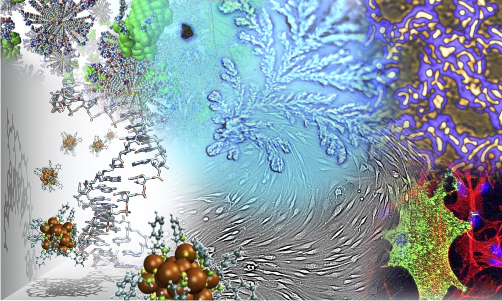

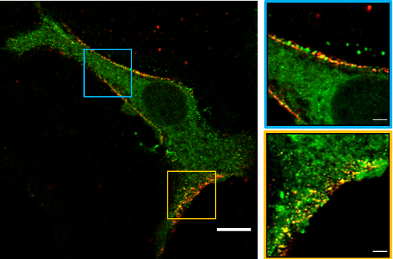
Fluorescence imaging is a straightforward and powerful method to disclose the interactions occurring between viruses and cells along the virus cycle from internalization to exocytosis. Yet, most viruses have a size around 100 nm, i.e. well below the optical resolution of conventional microscopes (200-300 nm). To address this issue, while maintaining the ability of fluorescence to afford a functional read-out of biomolecular interplays, we set up a multi-scale microscopy imaging toolbox leveraging both confocal and super-resolution microscopy.
The latter approach includes scanning techniques such as Image Scanning Microscopy (ISM) and Stimulated Emission Depletion (STED) microscopy, which can be applied also to living specimens from 120 to 60 nm, as well as Single Molecule Localization Microscopy (SMLM), which can reach 10-20 nm in fixed samples. Our toolbox is currently applied in the context of SARS-CoV-2 infection, and we recently revealed molecular hallmarks of virus “late entry” cell uptake mechanism, attributing a major role to clathrin-mediated endocytosis. Current activity focuses on the virus-cell receptor interaction at the cell membrane and how the internalization pathway is activated
| People | Ranieri Bizzarri, Barbara Storti* |
| Keywords | Viruses, virus-cell interactions, super-resolution fluorescence microscopy, endocytosis, virus-cell receptor interplay |
| Methods, techniques | ISM, Image Scanning Microscopy; SMLM, Single Molecule Localization Microscopy; STED, STimulated Emission Depletion; STORM, STochastic Optical Reconstruction Microscopy; TIRF, Total Internal Reflection Fluorescence; cell cultures; immunostaining |
| Collaborations | Prof. Mario Pistello and Paola Quaranta,Retrovirus Center, Department of Translational Research and New Technologies in Medicine and Surgery, University of Pisa (IT) and Pisa University Hospital; Nicola Clementi and Elena Criscuolo, Laboratory of Medical Microbiology and Virology, University “Vita-Salute” San Raffaele and Laboratory of Medical Microbiology and Virology, IRCCS San Raffaele Hospital, Milan, Italy; Gianmarco Ferri, Fondazione Pisana per la Scienza, Pisa (IT); Alberto Diaspro, Nanoscopy, CHT, Istituto Italiano di Tecnologia, Genoa (IT); Margherita Maffei, CNR-IFC, Pisa |
| Publications | |
| B. Storti, P. Quaranta, C. Di Primio, N. Clementi, N. Mancini, E. Criscuolo, P. G. Spezia, V. Carnicelli, G. Lottini, E, Paolini, G. Freer, M. Lai, M. Costa, F Beltram, A. Diaspro, M. Pistello, R. Zucchi, P. Bianchini, G. Signore and R. Bizzarri A spatial multi-scale fluorescence microscopy toolbox discloses entry checkpoints of SARS-CoV-2 variants in Vero E6 cells. Comput Struct Biotechnol J. 2021 | |
| F Saponaro, G Rutigliano, S Sestito, L Bandini, B Storti, R Bizzarri, R Zucchi ACE2 in the era of SARS-CoV-2: controversies and novel perspectives Front. Mol. Biosci. 2020 |